Stanford transplants human brain organoids into cortex-less mice: after three months, 90% of the cortex was human
A host that lacks a cortex
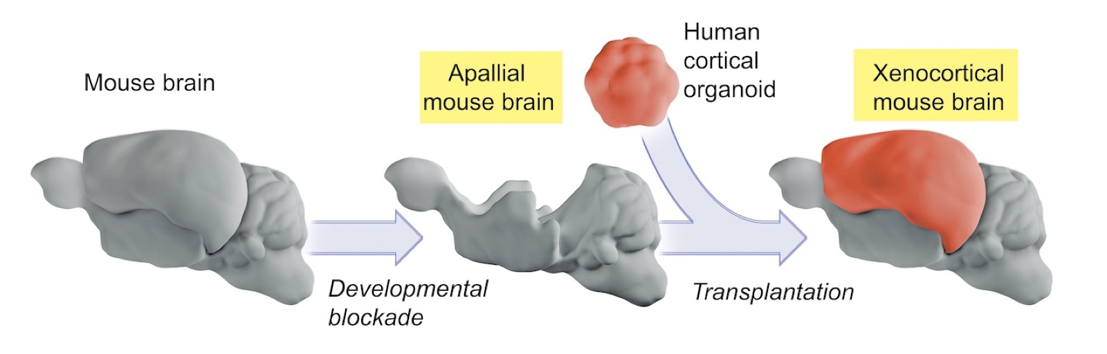
On 16 September Nature published online a study from Stanford Medicine: the team transplanted laboratory-grown human cortical organoids — self-organizing three-dimensional pieces of brain tissue — into mice engineered to lack almost their entire cerebral cortex. The cerebral cortex is the outermost rind of the brain, credited with much of our higher-level functioning such as cognition, language, attention and decision-making.
The work is worth doing because severe neurodevelopmental disorders are exceedingly difficult to study at the molecular and cellular level: the brain is more complex than any other organ, and living human brain tissue is nearly always inaccessible. By Stanford's numbers, one in 20 American adults is troubled by a severe psychiatric illness; more than 1 in 100 adults has schizophrenia, believed to result largely from brain-circuit abnormalities predating birth; and about 1% of people remain epileptic throughout adulthood. Alison Singer, president of the Autism Science Foundation, gives another figure: one in every 218 American children meets the criteria for profound autism, with measured IQs below 50, requiring round-the-clock supervision.
"These animal models offer a unique opportunity to study how disease-associated alterations in human brain circuitry manifest in an intact nervous system," says Sergiu Pasca, the study's senior author and director of the Stanford Brain Organogenesis Program. "Neuroscientists will be able to learn much more about the causes and mechanisms of neurodevelopmental and pregnancy-incurred disorders and to test possible interventions to correct or prevent them," he adds.
Three months later, more than 90% of the cortex was human
The road took more than a decade. The team first turned human skin cells into stem cells that can differentiate into most of the body's cell types, then coaxed them into self-organizing clusters of brain tissue whose internal architecture closely resembles cortical circuitry — what are now called organoids. They then juxtaposed organoids mimicking distinct brain regions so that their neurons interpenetrate and form complex signalling circuits; Pasca named these fused combinations assembloids, and three or four can be connected to recreate pathways that drive movement or sensation. Pasca says the team can now generate a broad range of brain cell types present throughout the developing human nervous system.
In 2022 they transplanted human cortical organoids into the brains of newborn rats: tissue measuring perhaps one-fifth of an inch across at transplantation occupied a full one-third of the hemisphere six months later, and at eight months the human neurons were substantially bigger, more electrically active and more elaborately branched than organoids generated the same way but left in a dish, and had integrated with rat neurons into working circuits. In the same study the team also transplanted tissue from patients with Timothy syndrome — a rare condition marked by severe autism and heightened vulnerability to epilepsy and schizophrenia — pinpointed the molecular defect and found a candidate drug that is now being prepared for phase 1/2 safety trials. Limits remained: a dish has no blood supply, no immune-cell infiltration, no sensory input and no motor output, and "we couldn't study complex human behavior in a dish"; in the rat brain, rat neurons developed much faster than human ones — "the two parallel developing systems are in competition for turf".
The new study's answer is a host with almost no competition. In several technically challenging steps the team generated mice that survived in healthy condition despite the absence of most of their cortex and hippocampus, a structure important for memory formation. By adulthood, the brains of these "apallial mice" contained a mere 2% of ordinary mice's equivalent cortical content, leaving plenty of space that human cortical cells could occupy.
At a glance the apallial mice are almost indistinguishable from normal mice, but they show minor, measurable behavioral quirks: a slightly more cautious gait, probably reflecting somewhat compromised fine-motor coordination, and a tendency to be more forgetful about recently encountered novel environments. Their behavioral performance also varied more from one individual to the next than normal controls did. Pasca suggests that when cortical circuitry is lost very early in development, the cortex may not be solely responsible for all the functions traditionally attributed to it, and other brain regions may compensate as the brain develops.
Surgery took place two days after the apallial mice were born. The organoids — derived from healthy donors who consented to having their cells transplanted into animals and containing perhaps 100,000 cells each — were positioned one or more per recipient in the enlarged fluid-filled cavities of the brain. A lopsided majority of these operations succeeded: the human tissue survived and thrived, and three months after surgery more than 90% by volume of the cortical tissue in the mice's brains was human, with human and mouse neurons integrating into circuits apace. Three to six months after transplantation, the xenocortical mice's performance on behavioral tests was generally similar to that of their same-age normal peers.
An unexpected appearance of von Economo neurons
Even more unexpected was a cell type. In the human tissue of the xenocortical mice the researchers detected von Economo neurons — nerve cells peculiar in several ways: previously observed only in postmortem brain tissue, never generated in culture, and absent from the earlier transplant experiments under more crowded conditions. They are very rare, accounting for roughly one in every 90,000 cortical neurons. Originally thought to be human-specific, they are now known to be present in other large-brained, highly social animals including great apes, elephants, dolphins and whales; in humans they sit in the fronto-insular cortex.
"This cell type appears to be particularly vulnerable in frontotemporal dementia," Pasca says. "In some forms of the disease, these cells are markedly reduced, and early symptoms can include changes in social behavior and personality or difficulties with language, often before prominent memory problems emerge." Von Economo neurons cannot be made in laboratory culture, he adds, "yet here they were, sitting in the xenocortical mice's human tissue. Now we can generate these rare cells from a healthy person and study them in a living, behaving animal to learn more about what they're doing. Or we can derive cells from patients with frontotemporal dementia or other neurological conditions, transfer them into these mice, test the animals for cellular or circuit-level correlates of behavioral disability, and then screen therapeutic candidates."
Oxygen deprivation, cerebral palsy, and an ethical boundary
The team also ran a proof-of-concept experiment showing how such mice can be used to study disease. Xenocortical mice were exposed to five hours of low oxygen, which wreaked substantial damage on their human-originated cortical tissue: the oxygen-deprived animals appeared to have trouble sustaining a steady gait and maintaining their balance, reminiscent of what is seen in children with cerebral palsy, while normal and apallial mice were virtually unaffected. Cerebral palsy affects 3 in 1,000 Americans and is a risk factor for epilepsy and autism. "Finding out what accounts for this difference could yield clues about human neural susceptibility to oxygen deprivation, shed light on mechanisms underlying cerebral palsy and provide a platform for testing potential therapeutic strategies," Pasca says.
Ethical questions run through the whole effort. Pasca stresses the importance of remaining within ethical boundaries: "Throughout several years of experimentation, we have received input from ethicists, neurobiologists with expertise in primate and human cortical biology, patient advocates, philosophers, and legal scholars." In November 2025 he organized a conference in Asilomar, California, to debate the ethical implications of using human stem cell models and their transplantation. He notes that one overriding argument questioned the ethics of not conducting this research in the face of the suffering of hundreds of millions of people afflicted with neurological disorders that today are incurable but tomorrow could yield treatments discovered with this model.
The boundaries deserve stating clearly too. This is an animal-model study: human tissue was transplanted into mice, and the mice remained mouse-like — the paper reports that their performance on behavioral tests was generally similar to that of normal peers, not that any human-like consciousness or cognition appeared. The real value is a research platform that did not exist before: a complete, living, experimentally accessible nervous system in which human brain circuitry and neurodevelopmental disease can be studied.